(IGP) IAS Pre: GS - Science & Technology : Biology - Lymphatic System and Immunity
Biology
Lymphatic System and Immunity
THE LYMPHATIC SYSTEM
-
The lymphatic system is composed of lymph vessels, lymph nodes, and organs. The functions of this system include the absorbtion of excess fluid and its return to the blood stream, absorption of fat (in the villi of the small intestine) and the immune system function.
-
Lymph vessels are closely associated with the circulatory system vessels. Larger lymph vessels are similar to veins. Lymph capillaries are scatted throughout the body. Contraction of skeletal muscle causes movement of the lymph fluid through valves.
- Lymph organs include the bone marrow, lymph nodes, spleen, and thymus.
- Bone marrow contains tissue that produces lymphocytes. B-lymphocytes (B-cells) mature in the bone marrow.
- T-lymphocytes (T-cells) mature in the thymus gland.
- Other blood cells such as monocytes and leukocytes are produced in the bone marrow.
- Lymph nodes are areas of concentrated lymphocytes and macrophages along the lymphatic veins.
- The spleen is similar to the lymph node except that it is larger and filled with blood.
- The spleen serves as a reservoir for blood, and filters or purifies the blood and lymph fluid that flows through it.
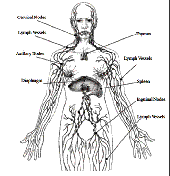
- If the spleen is damaged or removed, the individual is more susceptible to infections.
- The thymus secretes a hormone, thymosin, that causes pre-T-cells to mature (in the thymus) into T-cells.
IMMUNITY
- Immunity is the body’s capability to repel foreign substances and cells.
- The nonspecific responses are the first line of defense.
- Highly specific responses are the second line of defense and are tailored to an individual threat.
- The immune response includes both specific and nonspecific components. Nonspecific responses block the entry and spread of disease-causing agents.
- Antibody-mediated and cell-mediated responses are two types of specific response.
- The immune system is associated with defense against disease-causing agents, problems in transplants and blood transfusions, and diseases resulting from over-reaction (autoimmune, allergies) and under- eaction (AIDS).
(A) GENERAL DEFENSES
Barriers to entry are the skin and mucous membranes.
-
The skin is a passive barrier to infectious agents such as bacteria and viruses. The organisms living on the skin surface are unable to penetrate the layers of dead skin at the surface. Tears and saliva secrete enzymes that breakdown bacterial cell walls. Skin glands secrete chemicals that retard the growth of bacteria.
-
Mucus membranes lining the respiratory, digestive, urinary, and reproductive tracts secrete mucus that forms another barrier. Physical barriers are the first line of defense.
-
When microorganisms penetrate skin or epitheliumlining respiratory, digestive, or urinary tracts, inflammation results. Damaged cells release chemical signals such as histamine that increase capillary blood flowinto the affected area (causing the areas to become heated and reddened). The heat makes the environment unfavorable for microbes, promotes healing, raises mobility of white blood cells, and increases themetabolic rate of nearby cells. Capillaries pass fluid into intelstinal areas, causing the infected/ injured area to swell.
-
Clotting factors trigger formation of many small blood clots. Finally, monocytes (a type of white blood cell) clean up dead microbes, cells, and debris.
-
If this is not enough to stop the invaders, the complement system and immune response act.
-
Protective proteins that are produced in the liver include the complement system of proteins. The complement system proteins bind to a bacterium and open pores in its membrane through which fluids and salt move, swelling and bursting the cell. The complement system directly kills microbes, supplements inflammatory response, and works with the immune response. It complements the actions of the immune system. Complement proteins are made in the liver and become active in a sequence (C1 activates C2, etc.). The final five proteins form a membraneattack complex (MAC) that embeds itself into the plasma membrane of the attacker.
-
Salts enter the invader, facilitating water to cross the membrane, swelling and bursting the microbe. Complement also functions in the immune response by tagging the outer surface of invaders for attack by phagocytes.
-
Interferon is a species-specific chemical produced by cells that are viral attack. It alerts nearby cells to prepare for a virus. The cells that have been contacted by interferon resist all viral attacks.
(B) SPECIFIC DEFENSES
- The immune system also generates specific responses to specific invaders.
- The immune system is more effective than the nonspecific methods, and has a memory component that improves response time when an invader of the same type (or species) is again encountered.
- Immunity results from the production of antibodies specific to a given antigen (antibody generators, located on the surface of an invader).
- Antibodies bind to the antigens on invaders and kill or inactivate them in several ways.
- Most antibodies are themselves proteins or are a mix of protein and polysaccharides. Antigens can be any molecule that causes antibody production.
Lymphocytes : White blood cells known as lymphocytes arise from mitosis of stem cells in the bone marrow. Some lymphocytes migrate to the thymus and become T cells that circulate in the blood and are associated with the lymph nodes and spleen.
B cells remain in the bone marrow develop before moving into the circulatory and lymph systems. B cells produce antibodies.
- Antibody-mediated (humoral) immunity is regulated by B cells and the antibodies they produce. Cell-mediated immunity is controlled by T cells.
- Antibody-mediated reactions defend against invading viruses and bacteria. Cell-mediated immunity concerns cells in the body that have been infected by viruses and bacteria, protect against parasites, fungi, and protozoans, and also kill cancerous body cells.
Antibody-mediated Immunity :
Stages in this process are :
- antigen detection
- activation of helper T cells
- antibody production by B cells
Each stage is directed by a specific cell type.
-
Macrophages : Macrophages are white blood cells that continually search for foreign (nonself) antigenic molecules, viruses, or microbes. When found, the macrophages engulfs and destroys them. Small fragments of the antigen are displayed on the outer surface of the macrophage plasma membrane.
-
Helper T Cells : Helper T cells are macrophages that become activated when they encounter the antigens now displayed on the macrophage surface. Activated T cells identify and activate B cells.
-
B Cells : B cells divide, forming plasma cells and B memory cells. Plasma cells make and release between 2000 and 20,000 antibody molecules per second into the blood for the next four or five days. B memory cells live for months or years, and are part of the immune memory system.
-
Antibodies : Antibodies bind to specific antigens in a lock-and-key fashion, forming an antigenantibody complex.Antibodies are a type of protein molecule known as immunoglobulins. There are five classes of immunoglobulins: IgG, IgA, IgD, IgE, and IgM.
Antibodies are Y-shaped molecules composed of two identical long polypeptide (Heavy or H chains) and two identical short polypeptides (Light or L chains). Function of antibodies includes:
- Recognition and binding to antigens
- Inactivation of the antigen
A unique antigenic determinant recognizes and binds to a site on the antigen, leading to the destruction of the antigen in several ways. The ends of the Y are the antigencombining site that is different for each antigen.
Helper T cells activate B cells that produce antibodies. Supressor T cells slow down and stop the immune response of B and T cells, serving as an off switch for the immune system. Cytotoxic (or killer) T cells destroy body cells infected with a virus or bacteria. Memory T cells remain in the body awaiting the reintroduction of the antigen.
A cell infected with a virus will display viral antigens on its plasma membrane. Killer T cells recognize the viral antigens and attach to that cell’s plasma membrane. The T cells secrete proteins that punch holes in the infected cell’s plasma membrane. The infected cell’s cytoplasm leaks out, the cell dies, and is removed by phagocytes. Killer T cells may also bind to cells of transplanted organs.
The immune system is the major component of this defense. Lymphocytes, monocytes, lymph organs, and lymph vessels make up the system. The immune system is able to distinguish self fromnon- elf.Antigens are chemicals on the surface of a cell. All cells have these. The immune system checks cells and identifies them as “self” or “nonself”. Antibodies are proteins produced by certain lymphocytes in response to a specific antigen. Blymphocytes and T-lymphocytes produce the antibodies. Blymphocytes become plasma cells which then generate antibodies. T-lymphocytes attack cells which bear antigens they recognize. They also mediate the immune response.

Click Here to Download full Chapter
Click Here for Lymphatic System & Immunity MCQ
THE IMMUNE SYSTEM AND MEMORY OF INFECTIONS
Secondary immunity, the resistance to certain diseases after having had them once, results from production of Memory B and T cells during the first exposure to the antigen. A second exposure to the same antigen produces a more massive and faster response. The secondary response is the basis for vaccination.
VACCINATION
Vaccination is a term derived from the Latin vacca (cow, after the cowpox material used by Edward Jenner in the first vaccination). A vaccine stimulates the antibody production and formation of memory cells without causing of the disease. Vaccines are made from killed pathogens or weakened strains that cause antibody production but not the disease. Recombinant DNA techniques can now be used to develop even safer vaccines. The immune system can develop long-term immunity to some diseases. Man can use this to develop vaccines, which produce induced immunity. Active immunity develops afteran illness or vaccine. Vaccines are weakened (or killed) viruses or bacteria that prompt the development of antibodies. Application of biotechnology allows development of vaccines that are the protein (antigen) which in no way can cause the disease. Passive immunity is the type of immunity when the individual is given antibodies to combat a specific disease. Passive immunity is short-lived.
BLOOD TYPES, RH, AND ANTIBODIES
There are 30 or more known antigens on the surface of blood cells. These form the blood groups or blood types. In a transfusion, the blood groups of the recipient and donor should match.
If improperly matched, the recipient’s immune system will produce antibodies causing clotting of the transfused cells, blocking circulation through capillaries and producing serious or even fatal results. Individuals with blood type ‘A’ have the A antigen on the surface of their red blood cells, and antibodies to type B blood in their plasma. People with blood type ‘B’ have the B antigen on their blood cells and antibodies against type A in their plasma.
Individualswith type ‘AB’ blood produce have antigens for Aand B on their cell surfaces and no antibodies for either blood type A or B in their plasma. Type O individuals have no antigens on their red blood cells but antigens of both A and B are in their plasma. People with type AB blood can receive blood of any type, So it is called as Universal Receptar. Those with type O blood can donate to anyone. So it is called as Universal Donor. Hemolytic disease of the newborn (HDN) results from Rh incompatibility between an Rh- mother and Rh+ fetus. Rh+ blood from the fetus enters the mother’s system during birth, causing her to produce Rh antibodies. The first child is usually not affected, however subsequent Rh+ fetuses will cause a massive secondary reaction of the maternal immune system.
To prevent HDN, Rh- mothers are given an Rh antibody during the first pregnancy with an Rh+ fetus and all subsequent Rh+ fetuses.
ORGAN TRANSPLANTS AND ANTIBODIES
Success of organ transplants and skin grafts requires a matching of his to compatibility antigens that occur on all cells in the body.
Chromosome 6 contains a cluster of genes known as the human leukocyte antigen complex (HLA) that are critical to the outcome of such procedures. The array of HLA alleles on either copy of our chromosome 6 is known as a haplotype.
The large number of alleles involved mean no two individuals, even in a family, will have the same identical haplotype.
Identical twins have a 100% HLA match. The best matches are going to occur within a family. The preference order for transplants is identical twin > sibling > parent > unrelated donor.
Chances of an unrelated donor matching the recipient range between 1 in 100,000-200,000.Matches across racial or ethnic lines are often more difficult. When HLA types are matched survival of transplanted organs dramatically increases.
DISORDERS OF THE IMMUNE SYSTEM
The immune system can overreact, causing allergies or autoimmune diseases. Likewise, a suppressed, absent, or destroyed immune system can also result in disease and death.
(a) Allergies : Allergies result from immune system hypersensitivity to weak antigens that do not cause an immune response in most people. Allergens, substances that cause allergies, include dust, molds, pollen, cat dander, certain foods, and some medicines (such as penicillin).
(b) Autoimmune diseases : The immune system usually distinguishes “self” from “nonself”. The immune systemlearns the difference between cells of the body and foreign invaders. Autoimmune diseases result when the immune system attacks and destroys cells and tissues of the body. Juvenile diabetes, Grave’s disease, Multiple sclerosis, Systemic lupus erythematosus, and Rheumatoid arthritis are some of the autoimmune diseases.
(c) Myasthenia gravis : Myasthenia gravis (MG) is a muscle weakness caused by destruction of muscle-nerve connections. Multiple sclerosis (MS) is caused by antibodies attacking the myelin of nerve cells. Systemic lupus erythematosis (SLE) has the person forming a series of antibodies to their own tissues, such as kidneys (the leading cause of death in SLE patients) and the DNA in their own cellular nuclei. In systemic lupus erythematosus (SLE), the immune system attacks connective tissues and major organs of the body.
(d) Immunodeficiency diseasesk : Immunodeficiency diseases result from the lack or failure of one or more parts of the immune system. Affected individuals are susceptible to diseases that normally would not bother most people. Genetic disorders, Hodgkin’s disease, cancer chemotherapy, and radiation therapy can cause immunodeficiency diseases.
(e) Severe Combined Immunodeficiency : Severe Combined Immunodeficiency (SCID) results froma complete absence of the cell-mediated and antibody-mediated immune responses. Affected individuals suffer froma series of seemingly minor infections and usually die at an early age. Asmall group suffering from adenosine deaminase (ADA) deficiency, a type of SCID, are undergoing gene therapy to provide them with normal copies of the defective gene.
(f) Acquired Immunodeficiency Syn-drome: Acquired Immunode-ficiency Syndrome (AIDS) is currently receiving the most attention among the immunodeficiency diseases. AIDS is a collection of disorders resulting from the destruction of T cells by the Human Immunodeficiency Virus (HIV), a retrovirus. When HIV replicates in the human T cells, it buds from the T cell plasma membrane encased in a coat derived from the T cell plasma membrane. HIV selectively infects and kills T4 helper cells. The viral RNA is converted into DNA by the enzyme reverse transcriptase; this DNA can become incorporated into a human chromosome for months or years. When the infected T cell is needed in the immune response, the viral genes are activated and the virus replicates, killing the infected cell and producing a new round on T4 cell infection. Gradually the number of T4 cells, the master on switch for the immune system, decline. The immune response grows less powerful, eventually failing. Premature death results from a series of rare diseases (such as fungal pneumonia and Kaposi’s sarcoma, a rare cancer) that overwhelm the body and its compromised immune system.
BODY DEFENCES
The specialised cells which deal with germs and forcing particles by eating them up are called ‘phagocytes’ (phagein ‘to eat’; cyte ‘cell’). They are present in all tissues but are particularly concentrated in liver, spleen and bone marrow.
- Monocyter in the blood are the circulating counterparts of these cells.
- Specific acquired immunity can be categorised into two groups: humoral immunity and cellular immunity
- Lymphoid organs produce lymphocytes. These organs include principally bone marrow, thymus, lymph, nodes, spleen and some ‘patches’ in the wall of the small intestine.
- The two types of lymphocytes— B lymphocytes concerned with humoral immunity, and T lymphocytes concerned with cellular immunity
- Antibody production takes place in Numoral immunity. It is triggered by a protein called the antigen. It is the plasma cells which manufacture antibodies specific for the antigen presented.
- Theories which sring to explain the synthesis of specific antibodies—‘in structure’ and ‘selective’ theories. Instructive thrones postulate that all plasma cells are alike, it is the antigen that directs the plasma cells to manufacture a specific protein (antibody)
- Selective theories originally proposed by Busnet, assume that there are as many types of B cells as the antigens Antibodies are proteins belonging to a class called ‘gamma globulins’ or immunoglobulins.
Hepatitis Vaccine— Three doses are required: the interval between the first and second dose being one month, and that between the second and third being six months. Oral typhoid vaccine is available in the formof capsule under the brand name ‘Typhoral’.
BLOOD: THE VITAL FLUID
Blood looks like a homogenous red fluid to the uncover edge. But when spread into a thin layer, it is found to be a suspension of different type of cells in a liquid called the ‘plasma’. Most of the cells are faint yellow and without a nucleus. A dense accumulation of these cells is responsible for the red colour of the blood. These cells are called ‘erythrocytes’ or red blood cells. These are also another two types of cells—the ‘leucocytes’ or white blood cells and ‘thrombocytes’ or platelets.
Plasma— is a straw coloured liquid, about 90 percent ofwhich iswater.The chief salt dissolved in plasma is sodium chloride, or common table salt. The salinity of plasma is onethird that of sea water.
- Fibrinogen is a protein which is essential for clotting of blood, another protein globulins aid in the defense mechanisms of the body.
- Red Blood Cells:– are the most numerous of the blood cells, they neither have a nucleus nor mitochondria, RBC are a reddish coloured protein containing iron.
-
It is hemoglobin whichmakes it possible to deliver oxygen to tissue which need it. The normal quantity of hemoglobin present in blood in 12-15 g in every 100 ml of blood. A decrease in this quantity is called ‘anemia’.
- The nucleus membrane of the roof of the mouth (palate) is the best region to access the quantity of hemoglobin.
- The average life span of a red cell is about four months. They are produced in the hollow of the bones (bone marrow).
-
White Blood Cells:–WBC are far less numerous than the RBC, the ratio being one white cell to every 600 red cells. They are slightly larger than the red cells, and differ in three aspects—first, they have nuclei, secondly, they do not contain hemoglobin, and are therefore nearly colourless, finally, some white cells can move and engulf particles or bacteria the process in called ‘phagocytosis’.
WBC are further subdivided in five groups.
- Neutroplis
- Eosinophils
- Basrophils
- Lymphocytes
- Monocytes
Platelets: are much smaller than red or white blood cells and are devoid of nuclei. They check the bleeding from an injury (harmostasis: haime ‘blood’; stages ‘standing’ Platelets contribute to this process of harmostasis by liberating a chemical called ‘sesolonies’.
- A, B, AB and O are the four blood groups. The classification is based on the type of substance present on the surface of red blood cells.
BLOOD ON THE MOVE: HEART AND BLOOD VESSELS
Blood move into the capillaries from the arteries. Arteries are large blood vessels which being the blood from the heart. Of the blood vessel, capillaries is the smallest and theirwalls the thinnest. It is the pulmonary veins which being book the oxygenated blood from the hungs to the heart.
- All arteries carry blood away from the heat. All veins bring blood back to the heat.
- All arteries except the pulmonary arteries, carry oxygenated blood, all veins except the pulmonary veins carry deoxygenated blood.
- The Heart is fist-sized, weight about 250 g and placed in the left side of the chest between the two hunge. It has right atrium and sign vent ride; left atrium and left vent ride.
The left ventride pumps the blood into the systematic circuit while the right ventricle pumps it into the pulmonary circuit. The first to work out this cycle was William Marvey.
-
The movement of blood within the heart is in accordance with the slope of pressure. The initial growth of blood from the left ventricle into the aorta gives rise to a wave which is transmitted to all the arteries. This wave is commonly looked for at the wrist and called the pulse. Each pulse heat corresponds to ventricular contraction, the rate at which the pulse is felt gives the heart rate.
- Congenital heart diseases are present sign at birth. They are due to defective formation of heart in the mothers numb.
- Babies having congenital heart disease are born ‘blue’ i.e. this lips, nail, as well as skin have a bluish finger.
Rheumatic heart disease:– usually starts as a sore throat. In rheumatic heart disease, generally the valves of the heart are involved.
Coronary Heart Disease:– generally affect individuals of a higher age group. Sometimes fatty substance are stuck of the inside coronary arteries, thereby reducing the supply of blood to the heart.
LUNGS: THE LIFE LINKE
The bronchial tree consists of larynx, trachea, bronchus left lung, right lung.
Alveoli – is a cluster of thin walled air sacs which end in tiny air cells. It is coveredwith a tracery of capillaries. A men has about 600 million alveoli.
- Oxygen move from the alveoli into the blood and carbondioxide move out of the capillaries to entre the alveoli.
FACTS FROM N.C.E.R.T
- Ball and Socket Joints:- The rounded end of one bone fits into the cavity (hollow space) of the other bone. Such a joint allows movements in all directions.
- Pivotal Joint:- The joint where our neck joins, the head is a pivotal joint. It allows us to bend our head forward and backward and turn the head to our right or left.
- Hinge Joint:- The elbow has a hinge joint that allows only a back and forth movement. Fixed Joint:- There are some bones in our head that are joined together at some joints the bones cannot move at these joints. Such joints are called fixed joints.
-
There is a joint between the upper jaw and the rest of the head which is a fixed joint. The ribs are curiously bent. They join the chest bone and the backbone together to forma box. This is called the rib cage.
- There are some additional parts of the skeleton that are not as hard as the bones and which can be bent. These are called cartilage
Click Here to Download full Chapter
Click Here for Lymphatic System & Immunity MCQ
© UPSCPORTAL.COM