(IGP) IAS Pre: GS - Science & Technology : Biology - The Nervous System
Biology
The Nervous System
INTRODUCTION
Multicellular animals must monitor and maintain a constant internal environment as well asmonitor and respond to an external environment. In many animals, these two functions are coordinated by two integrated and coordinated organ systems: the nervous systemand the endocrine system. Three basic functions performed by nervous systems are:-
- Receive sensory input from internal and external environments
- Integrate the input
- Respond to stimuli
SENSORY INPUT
Receptors are parts of the nervous system that sense changes in the internal or external environments. Sensory input can be in many forms, including pressure, taste, sound, light, blood pH, or hormone levels, that are converted to a signal and sent to the brain or spinal cord.
INTEGRATION AND OUTPUT
In the sensory centers of the brain or in the spinal cord, the barrage of input is integrated and a response is generated. The response, a motor output, is a signal transmitted to organs than can convert the signal into some form of action, such as movement, changes in heart rate, release of hormones, etc.
ENDOCRINE SYSTEMS
Some animals have a second control system, the endocrine system. The nervous system coordinates rapid responses to external stimuli. The endocrine systemcontrols slower, longer lasting responses to internal stimuli. Activity of both systems is integrated.
DIVISIONS OF THE NERVOUS SYSTEM
- The nervous system monitors and controls almost every organ system through a series of positive and negative feedback loops.
- The Central Nervous System (CNS) includes the brain and spinal cord.
- The Peripheral Nervous System (PNS) connects the CNS to other parts of the body, and is composed of nerves (bundles of neurons).
- Not all animals have highly specialized nervous systems.
- Those with simple systems tend to be either small and very mobile or large and immobile.
- Large, mobile animals have highly developed nervous systems: the evolution of nervous systems must have been an important adaptation in the evolution of body size and mobility.
NERVOUS SYSTEM IN VARIOUS ORGANISMS
Coelenterates, cnidarians, and echinoderms have their neurons organized into a nerve net. These creatures have radial symmetry and lack a head. Although lacking a brain or either nervous system (CNS or PNS) nerve nets are capable of some complex behavior. Bilaterally symmetrical animals have a body plan that includes a defined head and a tail region. Development of bilateral symmetry is associated with cephalization, the development of a head with the accumulation of sensory organs at the front end of the organism. Flatworms have neurons associated into clusters known as ganglia, which in turn form a small brain. Vertebrates have a spinal cord in addition to a more developed brain. Chordates have a dorsal rather than ventral nervous system. Several evolutionary trends occur in chordates: spinal cord, continuation of cephalization in the form of larger and more complex brains, and development of a more elaborate nervous system.
VERTEBRATE NERVOUS SYSTEM
The vertebrate nervous system is divided into a number of parts. The central nervous system includes the brain and spinal cord. The peripheral nervous system consists of all body nerves. Motor neuron pathways are of two types: somatic (skeletal) and autonomic (smoothmuscle, cardiac muscle, and glands).The autonomic system is subdivided into the sympathetic and parasympathetic systems.
-
Peripheral Nervous System The Peripheral Nervous System (PNS) contains only nerves and connects the brain and spinal cord (CNS) to the rest of the body. The axons and dendrites are surrounded by a white myelin sheath. Cell bodies are in the central nervous system (CNS) or ganglia. Ganglia are collections of nerve cell bodies. Cranial nerves in the PNS take impulses to and from the brain (CNS). Spinal nerves take impulses to and away from the spinal cord. There are two major subdivisions of the PNS motor pathways: the somatic and the autonomic.
Two main components of the PNS:
- sensory (afferent) pathways that provide input from the body into the CNS.
- motor (efferent) pathways that carry signals to muscles and glands (effectors).
Most sensory input carried in the PNS remains below the level of conscious awareness. Input that does reach the conscious level contributes to perception of our external environment.
A. SOMATIC NERVOUS SYSTEM
The Somatic Nervous System (SNS) includes all nerves controlling the muscular systemand external sensory receptors. External sense organs (including skin) are receptors. Muscle fibers and gland cells are effectors.
The reflex arc is an automatic, involuntary reaction to a stimulus. When the doctor taps your knee with the rubber hammer, she/he is testing your reflex (or knee-jerk). The reaction to the stimulus is involuntary, with the CNS being informed but not consciously controlling the response.
Examples of reflex arcs include balance, the blinking reflex, and the stretch reflex.Sensory input from the PNS is processed by the CNS and responses are sent by the PNS from the CNS to the organs of the body. Motor neurons of the somatic system are distinct from those of the autonomic system. Inhibitory signals, cannot be sent through the motor neurons of the somatic system.
B. AUTONOMIC NERVOUS SYSTEM
The Autonomic Nervous System is that part of PNS consisting of motor neurons that control internal organs. It has two subsystems. The autonomic system controls muscles in the heart, the smooth muscle in internal organs such as the intestine, bladder, and uterus. The Sympathetic Nervous System is involved in the fight or flight response. The Parasympathetic Nervous System is involved in relaxation. Each of these subsystems operates in the reverse of the other (antagonism).
Both systems innervate the same organs and act in opposition to maintain homeostasis. For example: when you are scared the sympathetic system causes your heart to beat faster; the parasympathetic system reverses this effect.
Motor neurons in this system do not reach their targets directly (as do those in the somatic system) but rather connect to a secondary motor neuron which in turn innervates the target organ.
Central Nervous System
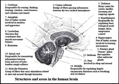
The Central Nervous System (CNS) is composed of the brain and spinal cord. The CNS is surrounded by boneskull and vertebrae. Fluid and tissue also insulate the brain and spinal cord.. The brain is composed of three parts: the cerebrum (seat of consciousness), the cerebellum, and the medulla oblongata (these latter two are “part of the unconscious brain.
The medulla oblongata is closest to the spinal cord, and is involved with the regulation of heartbeat, breathing, vasoconstriction (blood pressure), and reflex centers for vomiting, coughing, sneezing, swallowing, and hiccuping. The hypothalamus regulates homeostasis. It has regulatory areas for thirst, hunger, body temperature, water balance, and blood pressure, and links the Nervous System to the Endocrine System. The midbrain and pons are also part of the unconscious brain. The thalamus serves as a central relay point for incoming nervous messages. The cerebellumis the second largest part of the brain, after the cerebrum. It functions for muscle coordination and maintains normal muscle tone and posture. The cerebellum coordinates balance.
The conscious brain includes the cerebral hemispheres, which are separated by the corpus callosum. In reptiles, birds, and mammals, the cerebrumcoordinates sensory data and motor functions.
The cerebrum governs intelligence and reasoning, learning andmemory.While the cause of memory is not yet definitely known, studies on slugs indicate learning is accompanied by a synapse decrease. Within the cell, learning involves change in gene regulation and increased ability to secrete transmitters.
a. The Brain
During embryonic development, the brain first forms as a tube, the anterior end ofwhich enlarges into three hollow swellings that form the brain, and the posterior of which develops into the spinal cord. Some parts of the brain have changed little during vertebrate evolutionary history.
VERTEBRATE EVOLUTIONARY TRENDS INCLUDE
- Increase in brain size relative to body size.
- Subdivision and increasing specialization of the forebrain, midbrain, and hindbrain.
- Growth in relative size of the forebrain, especially the cerebrum, which is associated with increasingly complex behavior in mammals.

Click Here to Download full Chapter
Click Here for The Nervous System MCQ
THE BRAIN STEM AND MIDBRAIN
The brain stemis the smallest and froman evolutionary viewpoint, the oldest and most primitive part of the brain. The brain stem is continuous with the spinal cord, and is composed of the parts of the hindbrain and midbrain. The medulla oblongata and pons control heart rate, constriction of blood vessels, digestion and respiration. The midbrain consists of connections between the hindbrain and forebrain. Mammals use this part of the brain only for eye reflexes.
THE CEREBELLUM
The cerebellum is the third part of the hindbrain, but it is not considered part of the brain stem. Functions of the cerebellum include fine motor coordination and body movement, posture, and balance. This region of the brain is enlarged in birds and controls muscle action needed for flight.
THE FOREBRAIN
The forebrain consists of the diencephalon and cerebrum. The thalamus and hypothalamus are the parts of the diencephalon. The thalamus acts as a switching center for nerve messages. The hypothalamus is a major homeostatic center having both nervous and endocrine functions.
The cerebrum, the largest part of the human brain, is divided into left and right hemispheres connected to each other by the corpus callosum. The hemispheres are covered by a thin layer of gray matter known as the cerebral cortex,themost recently evolved region of the vertebrate brain. Fish have no cerebral cortex, amphibians and reptiles have only rudiments of this area.
The cortex in each hemisphere of the cerebrum is between 1 and 4 mm thick. Folds divide the cortex into four lobes: occipital, temporal, parietal, and frontal. No region of the brain functions alone, although major functions of various parts of the lobes have been determined.
The occipital lobe (back of the head) receives and processes visual information. The temporal lobe receives auditory signals, processing language and the meaning of words. The parietal lobe is associated with the sensory cortex and processes information about touch, taste, pressure, pain, heat and cold. The frontal lobe conducts three functions:
- motor activity and integration of muscle activity
- speech
- thought processes
Most peoplewho have been studied have their language and speech areas on the left hemisphere of their brain. Language comprehension is found in Wernicke’s area. Speaking ability is in Broca’s area. Damage to Broca’s area causes speech impairment but not impairment of language comprehension. Lesions in Wernicke’s area impairs ability to comprehend written and spoken words but not speech. The remaining parts of the cortex are associated with higher thought processes, planning, memory, personality and other human activities.
b. The Spinal Cord
The spinal cord runs along the dorsal side of the body and links the brain to the rest of the body. Vertebrates have their spinal cords encased in a series of (usually) bony vertebrae that comprise the vertebral column. The gray matter of the spinal cord consists mostly of cell bodies and dendrites. The surrounding white matter is made up of bundles of interneuronal axons (tracts). Some tracts are ascending (carrying messages to the brain), others are descending (carrying messages from the brain). The spinal cord is also involved in reflexes that do not immediately involve the brain.
NEUROLOGICAL DISEASES
Neurological diseases, for example Parkinson’s disease and Huntington’s disease, are due to imbalances of neurotransmitters. Parkinson’s is due to a dopamine deficiency. Huntington’s disease is thought to be cause by malfunctioning of an inhibitory neurotransmitter. Alzheimer’s disease is associated with protein plaques in the brain.
THE NEURON
Nervous tissue is composed of two main cell types: Neurons and glial cells. Neurons transmit nerve messages. Glial cells are in direct contact with neurons and often surround them. The neuron is the functional unit of the nervous system. Humans have about 100 billion neurons in their brain alone!While variable in size and shape,
a. Parts of Neuron:
All neurons have three parts.
Dendrites receive information from another cell and transmit the message to the cell body.
The cell body contains the nucleus, mitochondria and other organelles typical of eukaryotic cells.
The axon conducts messages away fromthe cell body.
b. Types of Neuron:
Three types of neurons occur. Sensory neurons typically have a long dendrite and short axon, and carry messages from sensory receptors to the central nervous system. Motor neurons have a long axon and short dendrites and transmit messages from the central nervous system to the muscles (or to glands).
Interneuron's are found only in the central nervous system where they connect neuron to neuron. Some axons are wrapped in a myelin sheath formed from the plasma membranes of specialized glial cells known as Schwann cells. Schwann cells serve as supportive, nutritive, and service facilities for neurons. The gap between Schwann cells is known as the node of Ranvier, and serves as points along the neuron for generating a signal. Signals jumping from node to node travel hundreds of times faster than signals traveling along the surface of the axon. This allows our brain to communicate with our toes in a few thousandths of a second.
THE NERVE MESSAGE
The plasma membrane of neurons, like all other cells, has an unequal distribution of ions and electrical charges between the two sides of the membrane. The outside of the membrane has a positive charge, inside has a negative charge.
Resting potential results from differences between sodium and potassium positively charged ions and negatively charged ions in the cytoplasm.
Sodium ions are more concentrated outside the membrane, while potassium ions are more concentrated inside the membrane. This imbalance is maintained by the active transport of ions to reset the membrane known as the sodium potassium pump. The sodium-potassium pump maintains this unequal concentration by actively transporting ions against their concentration gradients.
The action potential begins at one spot on the membrane, but spreads to adjacent areas of the membrane, propagating the message along the length of the cell membrane. After passage of the action potential, there is a brief period, the refractory period, during which the membrane cannot be stimulated. This prevents the message from being transmitted backward along the membrane.
STEPS IN AN ACTION POTENTIAL
- At rest the outside of the membrane is more positive than the inside.
- Sodium moves inside the cell causing an action potential, the influx of positive so diumionsmakes the inside of the membrane more positive than the outside.
- Potassium ions flow out of the cell, restoring the resting potential net charges.
- Sodium ions are pumped out of the cell and potassium ions are pumped into the cell, restoring the original distribution of ions.
SYNAPSES
The junction between a nerve cell and another cell is called a synapse. Messages travel within the neuron as an electrical action potential. The space between two cells is known as the synaptic cleft. To cross the synaptic cleft requires the actions of neurotransmitters. Neurotransmitters are stored in small synaptic vessicles clustered at the tip of the axon. Neurotransmitters tend to be small molecules, some are even hormones. The neurotransmitters cross the cleft, binding to receptor molecules on the next cell, prompting transmission of the message along that cell’s membrane. Diseases that affect the function of signal transmission can have serious consequences. Parkinson’s disease has a deficiency of the neurotransmitter dopamine. Progressive death of brain cells increases this deficit, causing tremors, rigidity and unstable posture.
SENSES
Input to the nervous system is in the form of our five senses: pain, vision, taste, smell, and hearing. Vision, taste, smell, and hearing input are the special senses. Pain, temperature, and pressure are known as somatic senses. Sensory input begins with sensors that react to stimuli in the formof energy that is transmitted into an action potential and sent to the CNS.
SENSORY RECEPTORS
- Sensory receptors are classified according to the type of energy they can detect and respond to.
- Mechanoreceptors: hearing and balance, stretching.
- Photoreceptors: light.
- Chemoreceptors: smell and taste mainly, as well as internal sensors in the digestive and circulatory systems.
- Thermoreceptors: changes in temperature.
- Electroreceptors: detect electrical currents in the surrounding environment.
Mechanoreceptors vary greatly in the specific type of stimulus and duration of stimulus/action potentials. Themost adaptable vertebrate mechanoreceptor is the hair cell. Hair cells are present in the lateral line of fish. In humans and mammals hair cells are involved with detection of sound and gravity and providing balance.
a. Hearing
Hearing involves the actions of the external ear, eardrum, ossicles, and cochlea. In hearing, sound waves in air are converted into vibrations of a liquid then into movement of hair cells in the cochlea. Finally they are converted into action potentials in a sensory dendrite connected to the auditory nerve. Very loud sounds can cause violent vibrations in the membrane under hair cells, causing a shearing or permanent distortion to the cells, resulting in permanent hearing loss.
Orientation and gravity are detected at the semicircular canals. Hair cells along three planes respond to shifts of liquid within the cochlea, providing a three-dimensional sense of equilibrium. Calciumcarbonate crystals can shift in response to gravity, providing sensory information about gravity and acceleration.
PHOTORECEPTORS DETECT VISION AND LIGHT SENSITIVITY
The human eye can detect light in the 400-700 nanometer (nm) range, a small portion of the electromagnetic spectrum, the visible light spectrum. Light with wavelengths shorter than 400 nm is termed ultraviolet (UV) light. Light withwavelengths longer than 700 nmis termed infrared (IR) light.
b. Eye
In the eye, two types of photoreceptor cells are clustered on the retina, or back portion of the eye. These receptors, rods and cones, apparently evolved from hair cells. Rods detect differences in light intensity; cones detect color. Rods are more common in a circular zone near the edge of the eye. Cones occur in the center (or fovea centralis) of the retina. Light reaching a photoreceptor causes the breakdown of the chemical rhodopsin, which in turn causes a membrane potential that is transmitted to an action potential. The action potential transfers to synapsed neurons that connect to the optic nerve. The optic nerve connects to the occipital lobe of the brain. Humans have three types of cones, each sensitive to a different color of light: red, blue and green. Opsins are chemicals that bind to cone cells and make those cells sensitive to light of a particular wavelength (or color). Humans have three different form of opsins coded for by three genes on the X chromosome. Defects in one or more of these opsin genes can cause color blindness, usually in males.
ANIMAL’S EYES
FACTS FROM NCERT:
- Eyes of a crab are quite small but they enable the crab to look all around.
- Butterflies have a large eye that seems to be made up of thousands of little eyes. They can see not only in the front and the sides but the back as well.
- Owl can see very well in the night but not during the day
- Kite, eagle can see well during the day but not in the night.
- The owl has large cornea and a large pupil to allow more light in its eye.Also it has on its retina a large number of rods and only a few cones.
- The day birds on the other hand have more cones and fewer rods.
- Louse Braille himself a visually challenged person developed a system for visually challenged persons and published it in 1821. Braille system has 63 dot pattern or characters.
- Diwaker a child prodigy has given amazing performances as a singer.
- Ravindra Jain showed his excellence as a lyricist, singer and music director.
- Lal advani established an association for special education and rehabilitation of disabled in India. He represented India on Braille problems in UNESCO.
- Hellen a kella is a famous American author and lecturer.
- Any person can donate their eyes suffering from any normal disease but not AIDS, hepatitis b or c, rabies, leukemia, tetanus, cholera, encephalitis.
- The eyes have to be donated within 4-6 hours after death.
HUMAN EYES AND A COLORFUL WORLD
To see an object comfortably we must hold it about 25 con from the eyes.
MYOPIA
- It is also known as short sightedness. A person with myopia can see nearby objects clearly but cannot see distinct objects distinctly.
- When light rays strike the retina, it stimulates highsensitive receptons called ‘rods’ ad ‘cones. Rods can perceive even dim light but cannot perceive a colour and do not give a very sharp image.
- Cones require bright light to be stimulated, respond differently to different colours, and give a very sharp image.
The sensitivity of rods and cones light is due to their containing a coloured substance called ‘pigment’. - Cones have three different types of pigments corresponding to these primary colours—green, blue and red.
Deficiency or absence of one or more type of cone pigments give rise to various focus of colour weakness or colour blindness.
Like haemophia, colour blindness is also a sex linked disease which affect only males.
Chemically all visual pigments are a combination of a derivative of Vitamin A and protein.
Click Here to Download full Chapter
Click Here for The Nervous System MCQ
© UPSCPORTAL.COM